What is Laparoscopic Living Donor Hepatectomy?
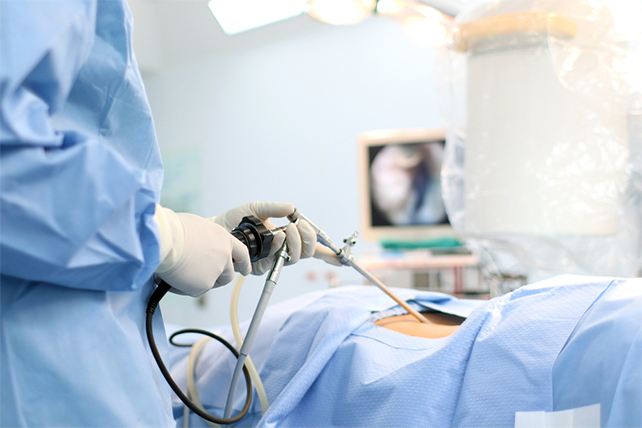
“Laparoscopic” surgery describes the techniques a surgeon uses to gain access to the internal surgery site. Most laparoscopic liver procedures start the same way. A laparoscope (a tiny telescope connected to a video camera) is inserted through the cannula, giving the laparoscopy Specialist a magnified view of the patient’s internal organs on a television monitor. A Technique known as Pure Laparoscopic Donor Hepatectomy allows surgeons to perform Laparoscopic living Donor hepatectomy through 4 or 5 small incisions (each about a quarter-inch). The liver graft is extracted through the lower abdomen scar (Pfannenstiel incision).
Details of the Surgery Given by Laparoscopy Specialist
For the operation to retrieve the donor graft, the patient was placed in the supine position with his legs apart and his head tilted up 15 degrees to help lower central venous blood pressure. The operating surgeon stood between the patient’s legs, with a view of the laparoscopic camera monitor placed in front of him, above the patient’s head.
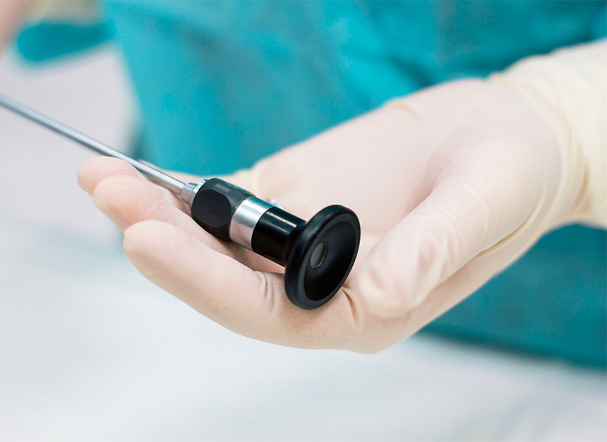
We prefer a 3-D scope instead of 2-D because it provides intuitive visual feedback with better depth perception. That may not be needed in simple surgeries such as cholecystectomy, but is essential in procedures as complex as living donor hepatectomy in order to perfect the operation.
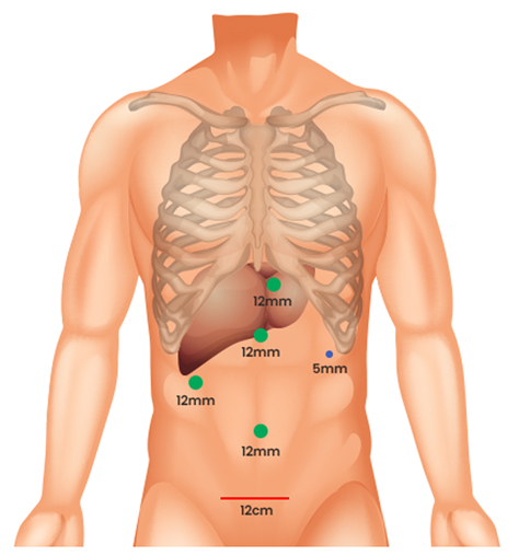
We placed five trocar ports: one 12mm at the umbilicus for optics; two 12 mm operative trocars on either side of the optics port, mainly for use by the operator; a 12 mm trocar below the sternum for retraction and exposure; and a 5 mm trocar in the left anterior axillary line.
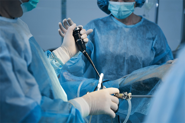
After mobilising the donor liver by dividing the triangular, falciform and coronary ligaments and performing a cholecystectomy, the best Laparoscopic Surgeon gently dissected and clamped the hepatic artery and portal vein.
With Laparoscopy Less Is More
 Less Post-Operative Pain
- Smaller Scars
- Quicker Return to solid food diet
- Less Recovery time
 Less Post-Operative Pain
 Smaller Scars
 Quicker Return to solid food diet
 Less Recovery time
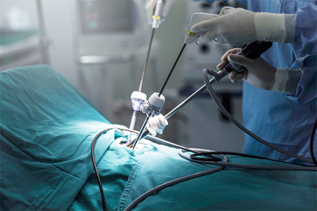
Fluorescent Intraoperative Indocyanine Green (ICG) Cholangiography was used for real-time visualisation, to delineate the left and right liver lobes for accurate parenchymal transection, and the bile duct anatomy for precise division. This advanced technique is based on the principle that intravenously injected ICG is excreted into bile and emits light when illuminated with a near-infrared light source attached to the laparoscopic camera. The best laparoscopic surgeon in Delhi can alternate between normal and near-infrared views on the camera monitor. After clamping, the differential in blood flow between the left and right liver creates a distinct demarcation line between the dark and light (fluorescing) halves, facilitating transaction. Likewise, bile duct anatomy is clearly illuminated. “With that fluorescence, you see the bile duct a lot better than when it’s not lit up,” says Doctor Vij.
After clamping, the differential in blood flow between the left and right liver creates a distinct demarcation line between the dark and light (fluorescing) halves, facilitating transaction. Likewise, bile duct anatomy is clearly illuminated. “With that fluorescence, you see the bile duct a lot better than when it’s not lit up,” says Doctor Vij . Following bile duct division and clipping, the remaining liver parenchymal transection was completed. He made a 9 cm Pfannenstiel incision in the donor’s abdomen to remove the graft, and inserted a plastic bag to contain it and aid handling. Placing the bag before dividing the hepatic artery reduces warm ischemia time. After injecting heparin 3,000 IU intravenously, he double-clipped and divided the hepatic artery. The portal and hepatic veins were divided and the remnant stumps were sealed with one-sided staples. We retrieved the graft through the incision, handed it off to the recipient team, then inserted drains and sutured the small abdominal opening.
Training of the Team by the Best Doctor for Laparoscopic Surgery
To prepare for the start of the AILBS laparoscopic LDLT program, the Best Doctor for Laparoscopic Surgery Dr Vij and his senior surgeons — each of whom has more than 10 years of experience in Open Oncological Liver surgery and living Donor Hepatectomies — operated together in liver resection cases, ultimately executing half of those cases laparoscopically. A dedicated team of scrub nurses and technicians was assembled, trained and took part in those procedures.
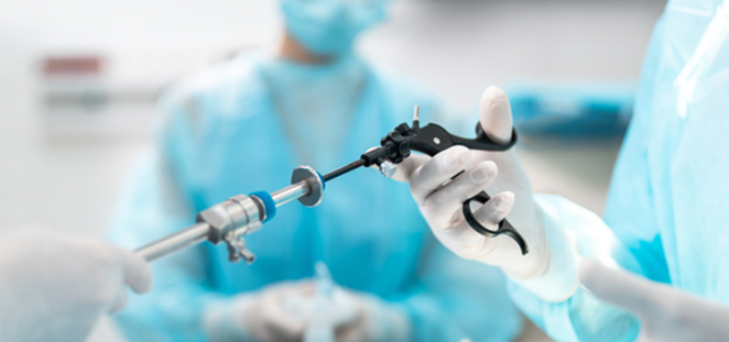
Essential Equipment Used by the Best Laparoscopic Surgeon
Advanced surgical equipment to help ensure optimal outcomes was purchased. The items include:


What Are The Benefits Of A Laparoscopic Operation For The Donor?
One of the main benefits of this procedure is that instead of an 8-20 inch scar that remains after open surgery, patients generally have a scar of about 3 inches. This scar is usually located in the lower abdomen, which tends to be an area that is less painful and less noticeable. In addition, we have observed reduced pain and shorter recovery times after surgery (less than 6 weeks, as opposed to 8-12 weeks for open surgery).
Common Advantages are:

May result in a faster return to a solid-food diet

May shorten hospital stay

Less postoperative pain

Quicker return to normal activity

May result in a quicker return of bowel function

Improved cosmetic results
What Complications can occur?

A bile leak where the liver was closed.

Injury to adjacent organs such as the small intestine, spleen, or pancreas

Blood clots to the lungs

Bleeding

Infection
Living Donor Hepatectomy: History, Benefits, Challenges & Success Story
A living Donor Hepatectomy is a procedure in which a portion of a healthy person’s liver is removed to be transplanted to a patient suffering from end-stage liver disease. Due to the liver’s unique ability to regenerate inside the body of both the donor and the recipient, only a portion of the liver is needed for a successful operation. While living donor hepatectomy has become quite common throughout India, AILBS team led by the Best Laparoscopic Surgeon in Delhi – Dr Vivek Vij remains the only centre performing this operation using minimally Invasive laparoscopic Technique in addition to standard open surgery.
Dr Vivek Vij is one of the world’s most experienced living donor liver transplant surgeons (laparoscopy specialist), having performed more than 1600 procedures at Fortis Hospitals, Delhi-NCR since founding that institution’s program in 2011. The procedure is technically demanding and is available only at a few hospitals worldwide, majorly in Asia. The laparoscopic surgery approach significantly reduces the invasiveness and morbidity of living donor liver procurement, improving safety for patients, shortening recovery times and potentially motivating more people to donate.
The Transition From Open To Laparoscopic Surgery
As the safety of altruistic donors is a priority due to significant morbidity and due to cosmetic concerns associated with open, large-incision of open liver donor surgery, some best laparoscopic surgeon began to contemplate whether the minimally- invasive laparoscopic surgery techniques employed in liver cancer resection and renal transplantation could be adapted for living-donor hepatectomy.
In 2002, a group of French surgeons was the first to show the feasibility of a laparoscopic approach for living liver donors, performing two successful parent-to-child transplants, involving left lateral sectionectomy. Laparoscopic living donor hepatectomy subsequently demonstrated advantages in reduced blood loss, postoperative morbidity, length of recovery and scarring compared to traditional open surgery. But the technique has not been widely adopted due to concerns about donor safety and a steep learning curve that requires expertise in both laparoscopic liver resection and LDLT.
“LDLT is one of the most complex surgeries that can be done laparoscopically,” Doctor Vij says. He is the best laparoscopic surgeon in Delhi who says “You need a lot of preparation. When we started the laparoscopic living liver donor program at AILBS , our team had already performed more than 1,500 LDLTs or laparoscopic liver transplants. That’s how we gained confidence and could manage the laparoscopic living- donor hepatectomy.”
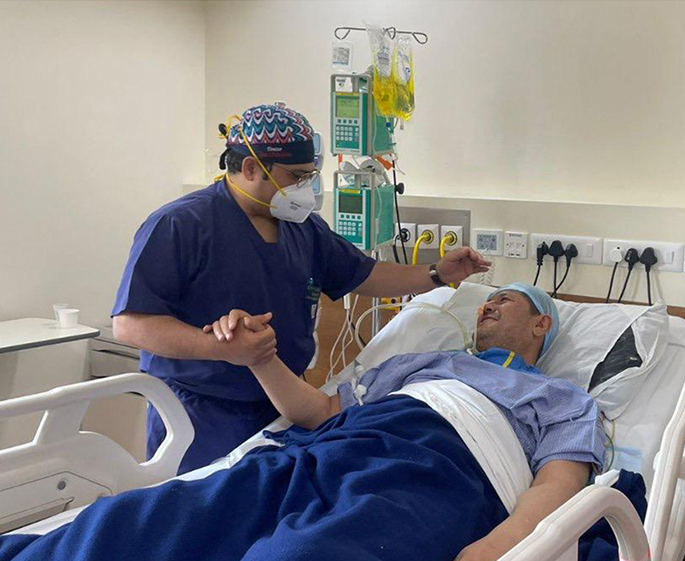
Success Stories
AILBS is the top liver transplant hospital in India, where Dr. Vivek Vij has performed thousands of liver transplant surgery. Let’s look at a few encouraging patient stories along the path to quality healthcare.
Frequently Asked Questions
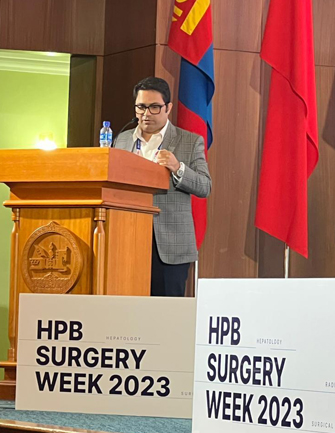
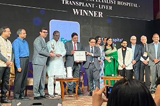
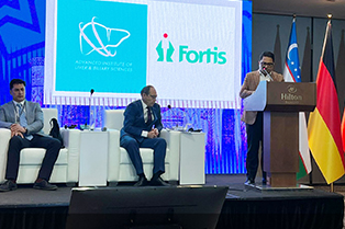
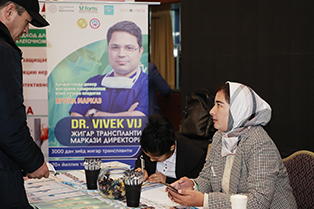
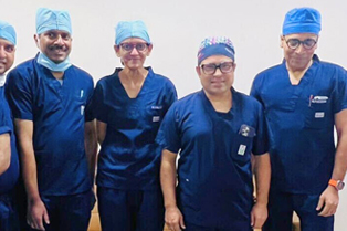
Gallery
Dr. Vivek Vij is a famous surgeon who is committed to providing compassionate and humane medical care to everyone who seeks the best care possible to have a healthy life. Let’s take a look at Dr. Vivek Vij, his knowledgeable staff, and his patients.

Gallery
Dr. Vivek Vij is a famous surgeon who is committed to providing compassionate and humane medical care to everyone who seeks the best care possible to have a healthy life. Let’s take a look at Dr. Vivek Vij, his knowledgeable staff, and his patients.